Describe How the Equipment Is Used in Mri
Having given up their energy the nuclei change their spin direction and return to the low energy state that they were in before. The human body is largely.
Mri Scans Definition Uses And Procedure
MRI equipment is essential to any major healthcare facility because it can create detailed images of the organs and tissues with the use of a strong magnetic field.

. Cooling the magnet to a temperature close to absolute zero 0 K allows such huge currents to be conducted. The modality makes it possible to scan any part of the body such as joints or abdomen within all direction. Those few leftover protons are the ones your MRI scanner will be using.
Radio frequency coils transmit the radio signal into the body part being imaged. Intra-ocular metallic foreign bodies intracranial aneurysm clips cardiac pacemakers and cochlear implants are generally counter-indicated for MRI. MRI scanners use strong magnetic fields magnetic field gradients and radio waves to generate images of the organs in the body.
Health care professionals use MRI scans to diagnose a variety of conditions from torn ligaments to tumors. It is often used for disease detection diagnosis and treatment monitoring. For imaging purposes the hydrogen nucleus a single proton is used because of its abundance in water and fat.
Receiver coils detect the returning radio signals. Newer MRI machines are open at the sides and of great benefit to claustrophobic patients. SIX MAIN COMPONENTS OF MRI SYSTEM Magnet Gradient Coils RF Coils Electronic Support System Computer Display 3 The magnet The magnet applies a static homogeneous magnetic field which align and precess the nuclei in the body.
Current diagnostic MRI scanners use cryogenic superconducting magnets in the range of 05 Tesla T to 15 T. Ad MRI Safe Products Maximize Safety Comfort For Both Patients Medical Staff. The hydrogen proton can be likened to the planet earth spinning on its axis with a north-south pole.
An MRI scanner contains two powerful magnets. Gradient coils provide spatial localization of the signals. Magnetic Resonance Imaging MRI is a non-invasive imaging technology that produces three dimensional detailed anatomical images.
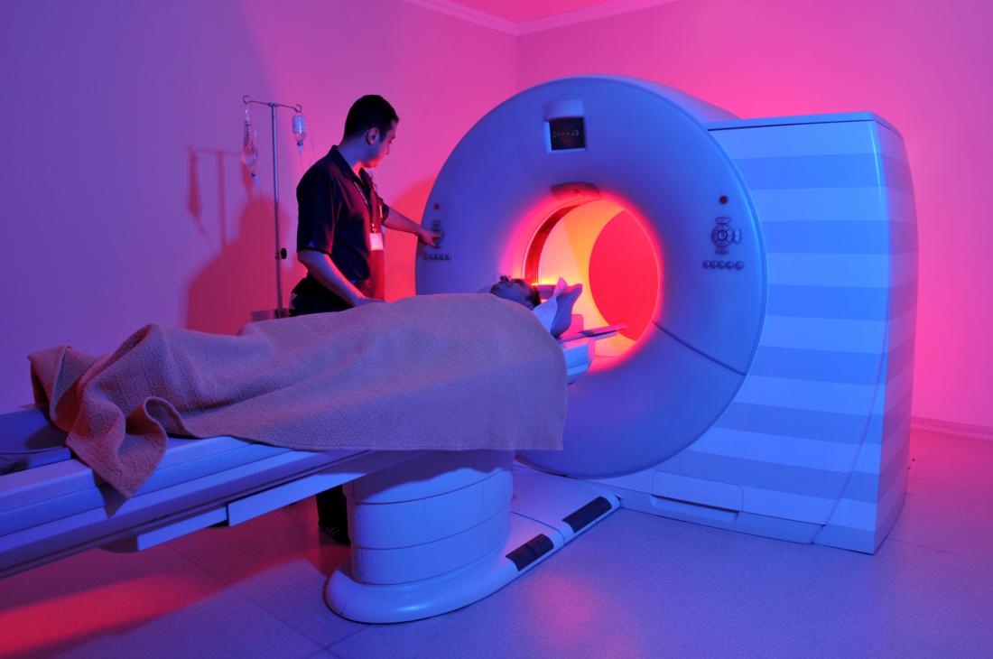
MRIs are very useful for examining the brain and spinal cord. During the scan you lie on a table that slides inside a tunnel-shaped machine. Ferromagnetic materials carry a projectile risk effect inside the MR scanner.
Also these scans are so clear from its high resolution in a way that differentiation of body tissues in a way that body components. This is most commonly performed via immersion in. By comparison the Earths magnetic field is 05 Gauss G which is equivalent to 000005 T.
These are the most important parts of the equipment. The major components are a computer system a magnet system a gradient system a radiofrequency system and a data acquisition system. The MRI machine has receiver coils blue coil shown below that receive the energy waves sent out by the nuclei.
It has advanced the field of diagnostic medicine to incredible heights. This coil is a radio transceiver that can communicate with your hydrogen atoms via radio frequency RF waves. What Kind of Equipment Is Used in an MRI.
The main advantage of the MRI over other imaging strategies is its accuracy in viewing in bones soft tissue and the blood vessels. A typical MRI machine is a large tube with a hole at both ends. The MRI equipment consists of following components.
MRI scans work by rearranging water molecules in the body with magnets. The DICOM standard provides a framework that allows. You may be gently strapped or secured in place to help prevent unintentional movement which may distort imaging.
This chapter describes the basic subsystems of an MRI scanner and technical aspects to consider when comparing scanners from different manufacturers. 2 4 Gradient Coils The gradient coils apply a variant of magnetic field strengths over the patient. CTs take a series of Xrays around a central axis either in a a discontinuous shoot and step process or b a continuous spiral manner When taken with b process is much quicker thus motion artefects are reduced which enables better 3D reconstruction of images.
Magnetic resonance imaging MRI uses a large magnet and radio waves to look at organs and structures inside your body. Every MRI patient has an RF coil placed near the part of the body being scanned. You are positioned on a padded and motorized table that slides in and out of the MRI tube or scanning area.
The receiver coil converts the energy waves into an electrical current signal. A magnet surrounds the tube. MRI does not involve X-rays or the use of ionizing radiation which distinguishes it from CT and PET scans.
The magnet generates the magnetic field. It is advisable to check their MR compatibility and for any counter-indications prior to the examination. The technologist uses that coil to send RF pulses at the body part under examination.
A magnetic resonance imaging instrument MRI scanner or nuclear magnetic resonance imaging scanner as it was originally known uses powerful magnets to polarize and excite hydrogen nuclei ie single protons of water molecules in human tissue producing a detectable signal which is spatially encoded resulting in images of the body. You lie on a. Shim coils make the magnetic field homogeneous.
By DirectMed Parts Service Aug 13 2021 MRI. Magnetic resonance imaging MRI uses the bodys natural magnetic properties to produce detailed images from any part of the body.
Introduction To Magnetic Resonance Imaging Mri

Magnetic Resonance Imaging Mri Has Been Described For The Past Few Decades As A Futuristic Imaging Technology That Will One Day Become Cardiac Mri Mri Study

Magnetic Resonance Imaging Mri


Comments
Post a Comment